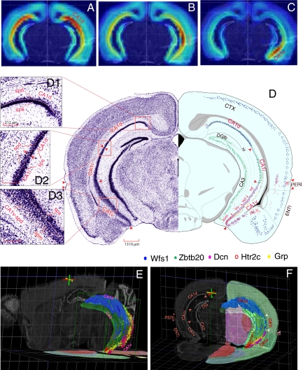
Fig. 1.
(A–C) Overall gene expression “heat maps” for “seed voxel” points in dorsal, intermediate, ventral domains of field CA1. (D) One transverse level of the ARA (15) displaying the maximal dorsoventral scope of field CA1 and its corresponding Nissl-stained histological images. Distributions of five marker genes, Wfs1 (blue), Zbtb20 (green), Dcn (purple), Htr2c (red), and Grp (yellow) are plotted on this level to reveal three molecular domains of field CA1 (CA1d, CA1i, and CA1v). (D1, D2, D3) High-resolution Nissl images that contain CA1d, CA1i, and CA1v, respectively (see text for details). (E) A three-dimensional model of Ammon's horn (in the context of the whole mouse brain). The overall shape of CA1 and its molecular domains revealed by CA1 gene markers Wfs1 (blue), Dcn (purple), and Htr2c (red) occupy the outside surface of the C-shaped cylinder of Ammon's horn, with the dorsal end (CA1d) extending much more rostral than the ventral end (CA1v). These two domains merge at the caudalmost end of CA1. (F) Three-dimensional expression patterns of 4 representative genes, Wfs1 (blue), Grp (yellow), Dcn (purple), and Htr2c (red), in one transverse plane of field CA1. These genes show distinct regional specificities and clearly define the spatial extent of CA1d (Wfs1), CA1v (Grp, Htr2c, and Dcn), and CA1i (revealed here by lack of these gene markers). Three-dimensional images of CA1 were generated in BrainExplore (http://www.brain-map.org), one three-dimensional model of the ARA (15, 40). See Fig. S1 for more detailed mapping of these genes in 4 representative levels of the ARA. CTX, cerebral cortex; DGlb, dentate gyrus, lateral blade; ENTl, lateral entorhinal area; PERI, perirhinal cortical area. (Scale bars: D, 1,310 μm; D1, D2, D3, 212 μm to match the same magnifications of the same images on ABA web site.)

