
Figure 3.
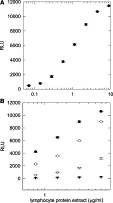
ELISA detection of FMRP from lymphocytes. ELISA analysis was performed on protein extracts from controls and fragile X syndrome patients. A: Sigmoidal dose-response curve of RLU (2.5 seconds light collection) as a function of protein concentration for control lymphocytes. Eight concentrations, from 0.2 to 6.0 μg/ml protein, were evaluated in triplicate. B: RLU values for (0.8 to 6.0 μg/ml protein) for two controls (filled circles, male; open circles, female) and two fragile X syndrome patients (triangles), all assayed in triplicate. Lymphocyte extract from the full-mutation male (filled triangles) showed minimal signal at all protein concentrations, whereas extract from a full-mutation female (open triangles; activation ratio, 0.46) yielded a detectable signal at higher concentrations.