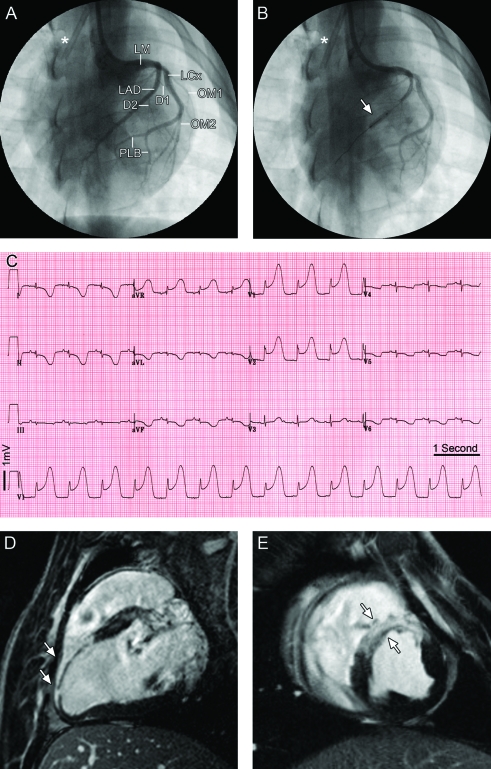
Figure 1.
Acute myocardial infarct in a Göttingen minipig. (A) Normal left coronary tree with branching of the anterior descending artery (LAD) and the left circumflex coronary artery (LCx) from the left main coronary artery (LM) in anterior-posterior projection. The LAD gives rise to 2 major diagonal branches (D1 and D2) supplying the anterior wall of the left ventricle. The left circumflex coronary artery (LCx) gives rise to 2 large obtuse marginal branches (OM1 and OM2), continues and ends in 2 posterior lateral branches (PLB). (B) Balloon inflation for temporary occlusion of the LAD after D2 (arrow). The asterisk (*) indicates a permanent central line placed in the superior vena cava. (C) The 12-lead ECG acquired 5 min after balloon occlusion shows the anterior-lateral location of the infarct with ST-segment depression in leads I, II, aVL, and aVF, and ‘tombstone’ ST elevation in the precordial leads. (D, E) Examples of delayed enhancement MRI of the same animal acquired on a 3-T magnet 3 d after MI to verify the anteroseptal location and demonstrate the transmurality of the induced MI (white arrows) along the (D) long axis and (E) short axis.

