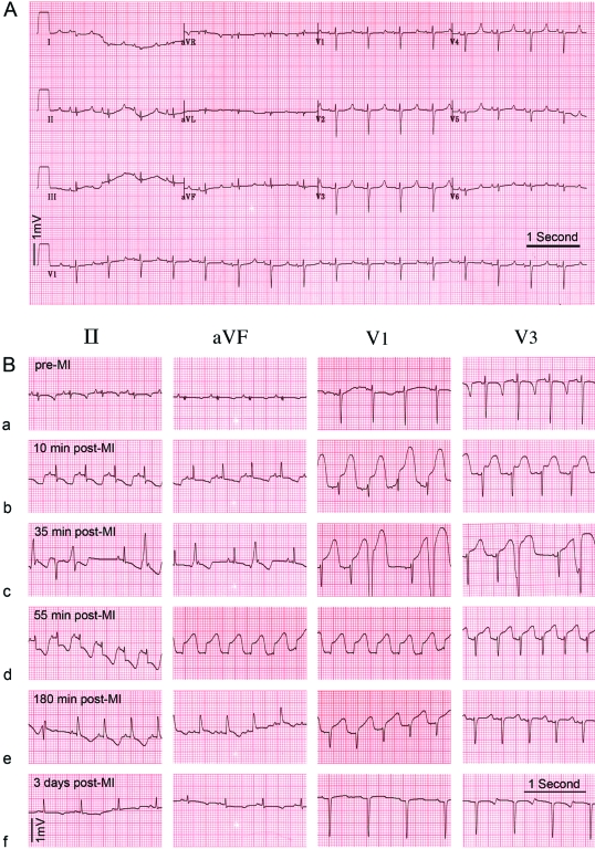
Figure 7.
Example of twelve-lead ECGs in Göttingen minipigs (A) Example of a normal ECG in an anesthetized Göttingen minipig (25 mm/s). (B) ECG time course in a Göttingen minipig during the MI procedure. (a) Baseline ECG prior to MI induction. (b) At 10 min after balloon occlusion, sinus tachycardia (123 beats per minute) with a short PR interval (100 ms) is present, with marked ST depression in II and aVF and ‘tombstone’ ST elevation in V1 and V3 (c) At 35 min into the MI period, frequent (>5 per minute) and consecutive premature ventricular complexes occurred. (d) At 55 min after balloon occlusion, the animal developed ventricular fibrillation. After cardioversion, the animal developed ectopic atrial tachycardia (162 beats per minute), which reverted to sinus tachycardia. (e) At 2.5 h after balloon occlusion, the infarct territory was reperfused. The ECG shows sign of transmural infarction after removal of the balloon catheter. (f) ST depression, which is most prominent in the aVF lead, is present 3 d after MI.

