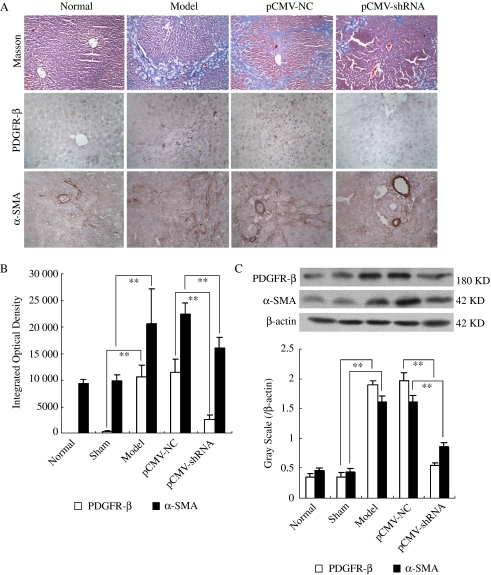
Fig. 6.
Effect of platelet-derived growth factor β subunit (PDGFR-β) short hairpin RNA (shRNA) on the histopathology and immunohistochemistry of experimental hepatic fibrosis induced by bile duct ligation (BDL). (A) Shows a representative graph of Masson's trichrome staining (the first line, magnification of × 20), PDGFR-β immunohistochemical staining (the second line, magnification of × 40) and α-smooth muscle actin (α-SMA) immunohistochemical staining (the third line, magnification of × 40). (B) Shows statistical results of PDGFR-β immunohistochemical staining and α-SMA immunohistochemical staining. (C) Shows representative graphs of PDGFR-β and α-SMA protein expression by Western blot analysis (up) and the statistical results (below). ‘Normal’ represents the normal group, ‘Sham’ represents the sham-operated group, ‘Model’ represents the model control group, ‘pCMV–NC’ represents the pCMV–NC–LacZ treatment group and ‘pCMV–shRNA’ represents the pCMV–shRNA–LacZ treatment group. Results show periportal fibrosis with short septa extending into lobules or porto–portal septa, severe cholestasis and bile duct hyperplasia in model control rats, while the hepatic fibrosis and cholestasis were all relieved by pCMV–shRNA–LacZ treatment. PDGFR-β and α-SMA protein expressions were significantly increased in model control rats, which was inhibited by pCMV–shRNA–LacZ treatment. **P<0.01.

