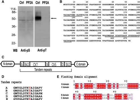
Figure 4.
Identification of MFP3 as the primary substrate for PP2A in fibers. (A) Western blot assays of MSP fibers obtained after treatment with KPM buffer (Ctrl) or with PP2A. Blots of duplicate gels were probed with anti-pS or anti-pT antibody. The arrow indicates the band that was excised from duplicate Coomassie-stained gels for peptide sequencing. (B) The deduced protein sequence of MFP3 from full-length cDNA, translated by proteomic tools at ExPasy (http://ca.expasy.org). Underlining shows the peptide sequences used for construction of primers for RT-PCR. (C) Diagram showing the domain organization of MFP3. Segments A–E are the tandem repeats, numeral indicate the numbers of each repeat. (D) Peptide compositions of tandem repeats in MFP3 sequence. Phosphorylation sites identified by FT-ICR MS/MS are highlighted in red. (E) The alignment of N-domain and C-domain of MFP3, which exhibited 61% amino acid identity.

