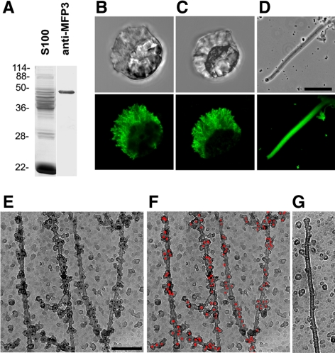
Figure 5.
MFP3 is a component of MSP motility apparatus. (A) Western blot and Coomassie-stained lanes of S100. The polyclonal antibody raised against MFP3 specifically recognized a single band. (B–D) Paired phase contrast and indirect immunofluorescence micrographs of a sperm probed with anti-MFP3 (B), a sperm labeled with anti-pT (C), and a fiber grown in vitro with anti-MFP3 (D). Bar, 10 μm. (E–G) Immunogold labeling of MSP filaments labeled with anti-MFP3 antibody and 10-nm colloidal gold particles coated with secondary antibodies. Gold particles are shown as black dots (E) and are highlighted in red pseudocolor in F. (G) A control MSP filament with the primary antibody omitted. Bar, 150 nm.

