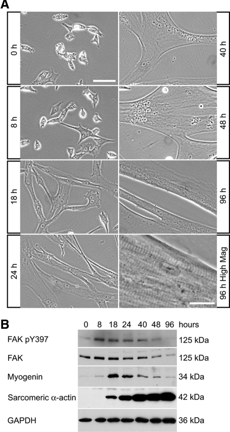
Figure 2.
Expression of FAK during myogenic differentiation in primary myoblast cultures. (A) Mouse primary myoblasts were cultured in DM and observed by phase contrast microscopy. Multinucleated myotubes formed within 48 h of initiation of differentiation. Myotube maturation is associated with the appearance of aligned sarcomeric striations and myotube contractions (seen at high magnification at 96 h). Bar, 100 μm (except for 96 h, high-magnification panel, 20 μm). (B) Primary myoblasts were cultured in DM then analyzed by Western blots. The levels of FAK and phosphorylated FAK were high during early differentiation then decreased as myotubes mature. Myogenin was transiently up-regulated during early differentiation, whereas sarcomeric α-actin increased dramatically with myogenic differentiation.

