Figure 6.
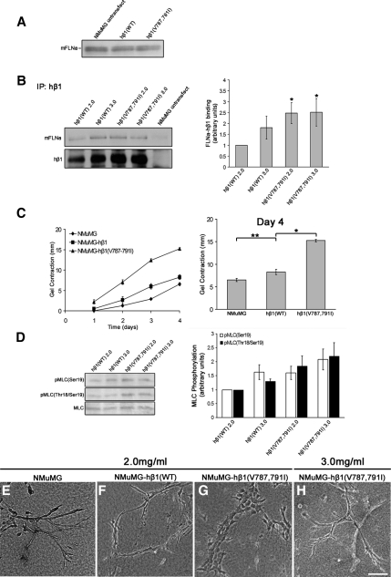
Increased FLNa binding to β1 integrin enhances matrix contraction and tunes branching morphogenesis. Mouse NMuMG cells were stably transfected with human β1 integrin wild-type (WT) or a β1 integrin containing two point mutations that specifically enhance binding of FLNa, hβ1(V787,791I). (A) Western blot of lysates from NMuMG cells expressing either hβ1(WT) or hβ1(V787,791I) demonstrates that these constructs did not alter levels of FLNa. Equal numbers of cells were lysed and used for comparison of mFLNa expression. (B) Cells expressing hβ1(V787,791I) cells have enhanced mFLNa binding to hβ1 integrin in both low- (2.0 mg/ml) and high (3.0 mg/ml)-density gels, whereas WT hβ1 integrin binding to mFLNa is regulated by matrix density. Immunoprecipitations (IPs) were performed with anti-hβ1 integrin antibody, the specificity of which was verified using equal numbers of untransfected NMuMG cells that do not express hβ1 integrin. Quantified data represent the mean ± SEM for four experiments. *p < 0.05; statistical difference compared with low-density control (two-sample t test). (C) Enhanced FLNa-β1 integrin interactions enhanced collagen gel contraction, shown over 4 d (left). Expression of hβ1(V787,791I) enhanced levels of gel contraction at day 4 relative to both hβ1(WT) and untransfected cells (right). Data are mean ± SEM from a minimum of eight experiments. *p < 0.001, **p < 0.05 statistical difference (two-sample t test). (D) Expression of hβ1(V787,791I) enhanced pMLC(Ser19) and pMLC(Thr18-Ser19) in both low- and high-density collagen gels. Quantified data represent the mean ± SEM for five experiments. p < 0.05 for all conditions; statistical significance relative to low-density hβ1 integrin control (two-sample t test). (E–H) Increased FLNa binding to β1 integrin tunes branching morphogenesis in high-density gels. Although NMuMG-hβ1(WT) cells exhibited a branched phenotype in low (2.0 mg/ml)-density gels (F), hβ1(V787–791I) cells exhibited disrupted morphogenesis in low-density gels (G). However, increasing the collagen density to 3.0 mg/ml was sufficient for hβ1(V787-791I) cells to undergo branching morphogenesis (H). Bar, 100 μm.

