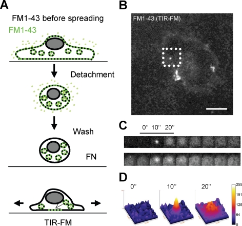
Figure 2.
Membrane exocytosis occurs during cell spreading. (A) Schematic representation of the FM1-43 before spreading protocol (see Materials and Methods). (B) Example of RPTPα cell TIR-FM images during spreading. Bar, 10 μm. (C) Sequential presentation of two vesicle fusions with the PM extracted from the Supplemental Video 1 (square selection from b is presented in the top row). (D) Fluorescence intensity surface plot analysis of the vesicle fusion presented in the top row of C. Height and colors range represents intensity.

