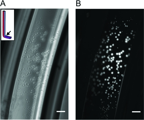
Figure 2.
Locally concentrated oleate forms vesicles in bent capillaries incubated at ΔT = 30 for 48 h. (A) Phase-contrast image of a bent capillary loaded with 70 μM buffered oleate and 40 μM HPTS. The oleate concentrated in the capillary and formed large vesicles. (B) Fluorescence image of the same frame. HPTS in the solution was washed away with dye-free buffer, leaving only encapsulated cargo to be visualized. Scale bars = 50 μm.

