Figure 1.
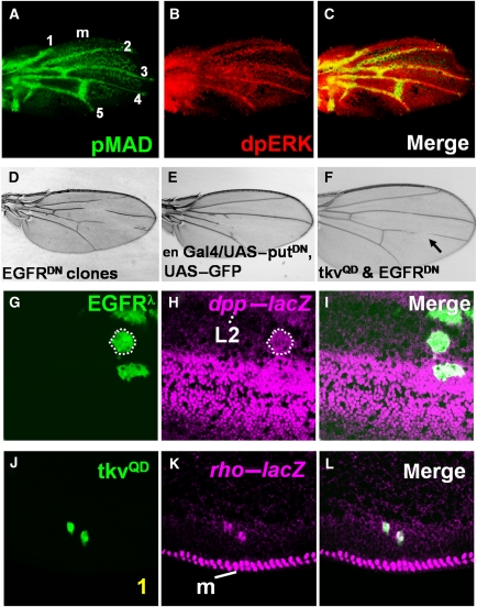
The DPP and EGFR pathways are jointly required for vein differentiation and reciprocally induce each other's ligands. (A–C) Pupal wings at 22 h APF stained for pMAD (green) and dpERK (red), with anterior (up) and distal (to the right), showing that both pMAD and dpERK are detected in the provein cells. 1–5, longitudinal veins L1–L5, and m, the wing margin. (D) Flip-out clones expressing EGFRDN-interrupted veins L2, L3, and L4. (E) Expressing PutΔI under control of the engrailed–Gal4 (en–Gal4) driver, which is expressed only in the posterior compartment of the wing, caused loss of vein tissues. (F) An adult wing with truncated veins resulting from co-expressing TKVQD and EGFRDN. In contrast to the wing with clones expressing TKVQD alone (Supplementary Figure S1A–C), few, if any, ectopic vein cells are observed (data not shown). (G–I) Ectopic EGFR signaling induces dpp expression. dpp–lacZ expression (magenta in H and I) is detected in the EGFRλ-expressing clones. (J–L) TKVQD activates EGFR by inducing rho expression.

