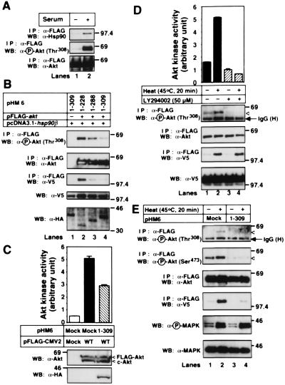
Figure 4.
Dephosphorylation and inactivation of Akt after detachment from Hsp90. (A) 293T cells were transfected with the FLAG-tagged WT-akt. After transfection for 24 h, cells were cultured in the medium containing no serum (−) or 10% FBS (+) for 24 h. Then, the FLAG-tagged Akt was immunoprecipitated and immunoblotted with the indicated antibodies. (B) The HA-tagged akt mutants, the V5-tagged WT-hsp90β, and/or the FLAG-tagged WT-akt were cotransfected into 293T cells. The cells were serum-starved for 24 h, and then FLAG-tagged WT-Akt was immunoprecipitated and immunoblotted with the indicated antibodies. The cell lysates were also immunoblotted with the indicated antibodies. (C) 293T cells were transfected with mock or the FLAG-tagged WT-akt together with mock or the HA-tagged 1–309 akt. The cells were serum-starved for 24 h and then harvested. The cell lysates were incubated with protein G agarose conjugated with a control IgG (lane 1) or an anti-Akt pAb (lanes 2 and 3). The Akt kinase activity of the immunoprecipitated Akt was estimated, as described in Materials and Methods. The vertical bars represent SD value of triplicate determinations. The immunoprecipitated proteins and the cell lysates were immunoblotted with the indicated antibodies. (D) HT1080 cells were transfected with the FLAG-tagged WT-akt together with V5-tagged WT-hsp90β. The cells were serum-starved for 24 h and then incubated at 37°C or 45°C for 20 min. In some experiments, cells were treated with 50 μM LY294002 for 10 min before heat shock. The Akt kinase activity of the immunoprecipitated FLAG-tagged Akt was estimated, as described in Materials and Methods. The vertical bars represent SD value of triplicate determinations. The cell lysates were also immunoblotted with the indicated antibodies. (E) HT1080 cells were transfected with the FLAG-tagged WT-akt, and the V5-tagged WT-hsp90β together with mock or the HA-tagged 1–309 akt. The cells were serum-starved for 24 h and then incubated at 37°C (−) or 45°C (+) for 20 min. The immunoprecipitated FLAG-tagged WT-Akt and the cell lysates were immunoblotted with the indicated antibodies. The expression of the HA-tagged 1–309 Akt was confirmed by immunoblot analysis with an anti-HA mAb (data not shown). Molecular size markers are indicated (in kDa).

