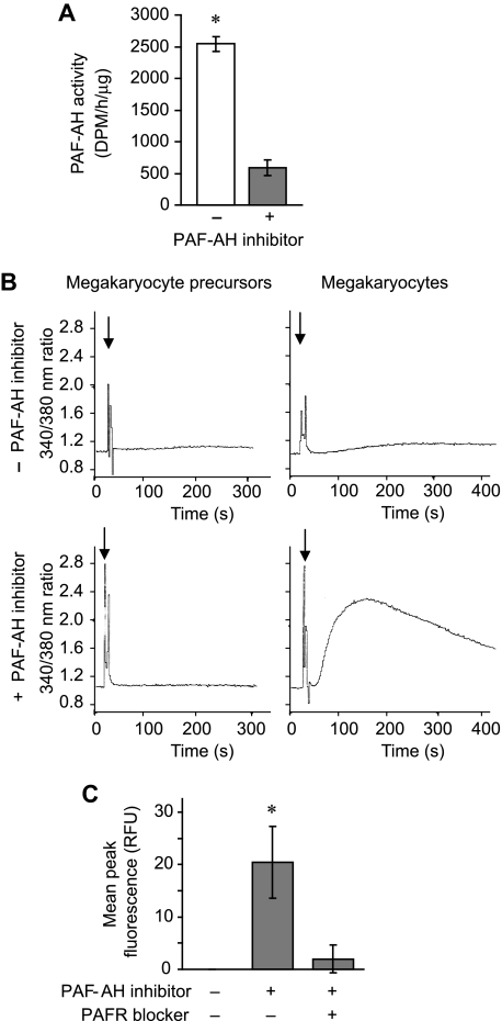
Figure 2.
Endogenous PAF-AH activity prevents megakaryocytes from accumulating phospholipids that activate the PAFR. (A) Intracellular PAF-AH activity (cell lysate) was measured as previously described32 in megakaryocytes adherent to immobilized fibrinogen in the presence or absence of 100 μM Pefabloc SC (PAF-AH inhibitor). The bars represent the mean ± SD for 3 independent experiments. (B) Megakaryocyte precursors (left column) or megakaryocytes (right column) were treated with vehicle (top row) or 100 μM of the PAF-AH inhibitor Pefabloc (bottom row). Phospholipids were extracted and added ( ) to Fura-2 AM–loaded human PMNs to measure intracellular calcium release by fluorometry. This figure is representative of 3 independent experiments. (C) Megakaryocytes adherent to immobilized fibrinogen were loaded with Fluo-4 AM in the presence or absence of the PAFR blocker, WEB 2086 (10 μM). The cells were subsequently treated with vehicle or 100 μM Pefabloc (PAF-AH inhibitor) to quench endogenous PAF-AH activity, and intracellular calcium fluxes were measured and are presented as mean peak fluorescence. The bars in panels A and C represent the mean ± SD for 3 independent experiments. * in panels A and C indicates statistical significance (P < .05) compared with untreated or other treatment groups.

