Figure 4.
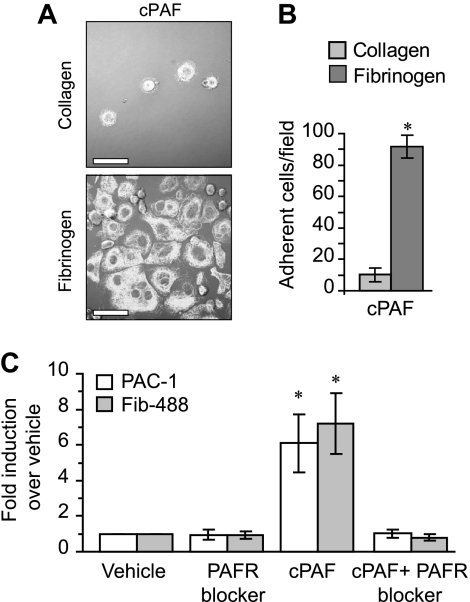
PAFR-dependent adherence and spreading in megakaryocytes relies on integrin αIIbβ3. (A) Megakaryocytes were placed on immobilized collagen or fibrinogen in the presence of 10 nM cPAF for 30 minutes. β-Tubulin was localized in the megakaryocytes (white stain). Scale bar represents 30 μm. (B) The number of megakaryocytes adherent to fibrinogen or collagen (ie, 30 minutes) in the presence of 10 nM cPAF. Panels A and B are representative of 3 independent experiments. (C) PAC-1 and Alexa 488–conjugated fibrinogen (Fib-488) staining in megakaryocytes that were left in suspension culture. The megakaryocytes were treated with 10 nM cPAF in the presence or absence of the PAFR blocker WEB 2086 (10 μM). The bars in panels B and C represent the mean ± SD. * identifies statistical significance (P < .05) between fibrinogen and collagen (B) or between cPAF and all the other treatments (C).

