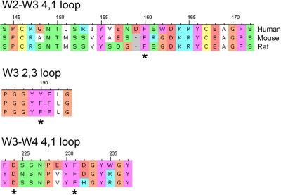
Figure 6.
Alignment of human, mouse, and rat αIIb sequences in the loops that contribute to the αIIb RGD ligand binding pocket. * indicates the 3 aromatic residues that line the pocket and the conserved D224 that interacts with the positively charged regions of the αIIbβ3 antagonists studied by X-ray crystallography.2 The nomenclature indicates the β-propeller blade number, and the loop designation identifies the β-sheets within the blades that are connected. The term loop is used broadly, since an α-helical region exists within the W2-W3 4,1 “loop.”

