Figure 5.
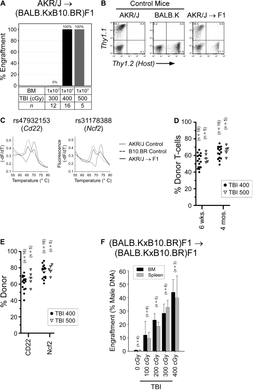
Allogeneic and syngeneic BM engraftment in (BALB.K × B10.BR)F1 mice. (A) F1 mice were given sublethal TBI and infused with AKR/J BM. Shown are the numbers of mice engrafting at 6 weeks after transplantation. For F1 mice engrafting with AKR/J BM, representative donor chimerism at 8 weeks after transplantation is shown with: (B) representative FACS analysis for T cells using Thy1.1 (donor) and Thy1.2 (host) cell-surface markers; (C) representative B-cell and myeloid chimerism by RT-PCR and melt curve analysis for Cd22 and Ncf2 coding SNP using total RNA extracted from the spleen of transplanted F1 recipient (solid line) or control mice (dotted and dashed lines); and dot plots summarizing (D) T-cell chimerism at indicated time points and (E) B-cell and myeloid chimerism at 4 months after transplantation. (F) Female F1 mice were given sublethal TBI and injected with syngeneic male BM. Shown are percent donor in BM (■) and spleen ( ) 6 weeks after transplantation by PCR for Sry DNA.

