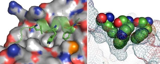
Fig. 1.

The X-ray crystal structure complex between a p53 peptide (residues 17–29; green CPK sticks and ribbon indicating secondary structure) and HDM2 (grey CPK surface). The three main p53 residue side chains (F19, W23, and L26) involved in the interaction are shown with solid sticks. Hydrogen bonds are indicated by dashed yellow lines (left). The p53-binding hot spot is illustrated on the right, where the three p53 residues are shown as a space-filled model, with the HDM2 surface as a mesh. Constructed from PDB # 1YCR. 3D structural illustrations were created using the programme PyMOL (DeLano, W.L. The PyMOL Molecular Graphics System (2002) DeLano Scientific, San Carlos, CA, USA. http://www.pymol.org).
