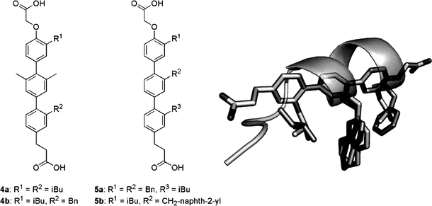
Fig. 7.

Substituted terphenyls 4 and 5 mimic the α-helical i, i+4, and i+7 side-chain arrangement found for the F19, W23, and L26 p53 residues (dark sticks and ribbon) in the HDM2-bound conformation. A likely conformation of terphenyl 5b (light sticks) was modelled and aligned with the three p53 residue side chains (PDB # 1YCR).
