
Figure 3. Deletions in the globular domain of CyPrP eliminate PrP’s anti-Bax function.
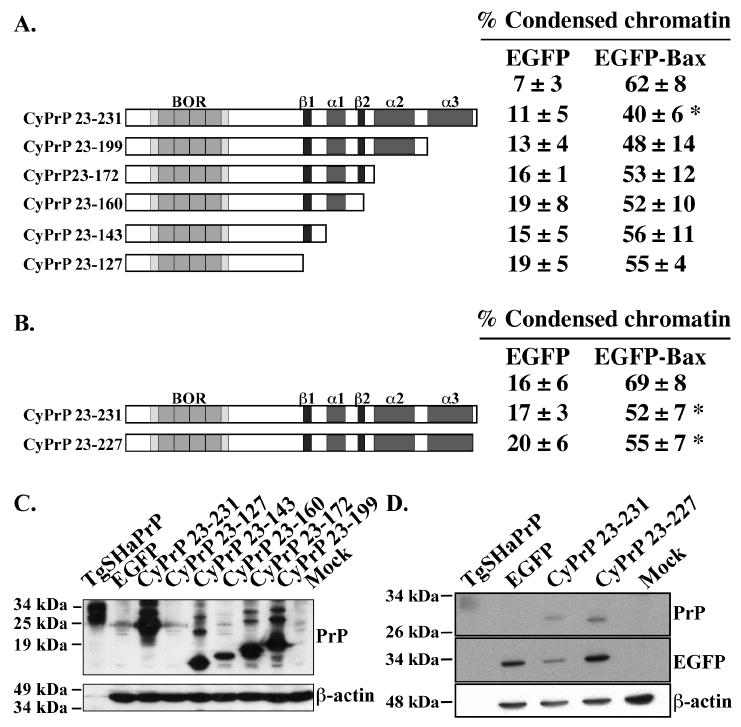
A & B. Schematic diagram of the sequential C-terminal CyPrP deletion mutants showing the main domains and structural elements. Right panel: Percentage (mean and SEM) of condensed chromatin positive cells in transfected MCF-7 cells. Data represents results from 4 to 5 independent experiments. At least 100 cells were counted for each experiment. Asterisk indicates a statistically significant difference from pBud-EGFP or pBud-EGFP-Bax (p≤0.05). C. Western blot analyses with 3F4 and β-actin antibodies of proteins extracted from cells transfected with CyPrP or CyPrP C-terminal globular domain deletion mutants. D. Western blot of total protein extracts from CyPrP23-231- or CyPrP23-227-transfected cells with 3F4, anti-GFP and anti-β-actin antibodies.