
Figure 4. Effect of CyPrP helix 3 mis-sense mutations on the anti-Bax function of PrP.
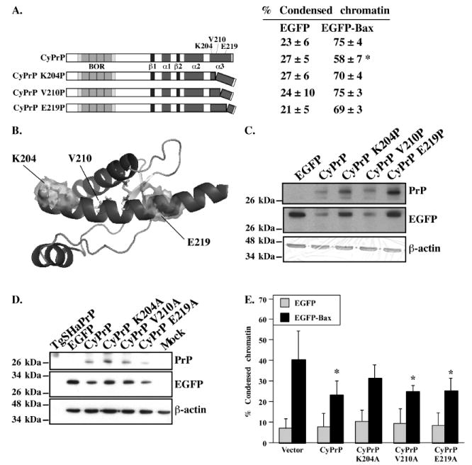
A. Schematic diagram of the CyPrP mutants with proline substitutions in helix 3.Right panel: Percentage (mean and SEM) of condensed chromatin positive cells in transfected MCF-7 cells. Data represents the results from 6 independent experiments. At least 100 cells were counted in each experiment. Asterisk indicates a statistically significant difference from pBud-EGFP or pBud-EGFP-Bax (p≤0.05). B. Location of the residues K204, V210 and E219 on the 3-dimensional structure of the CyPrP globular domain. C and D. Western blot analyses in protein extracts from cells transfected with CyPrP, the CyPrP proline substitution mutants (C) or the CyPrP alanine substitution mutants (D) with 3F4, anti-GFP or anti-β-actin antibodies. E. Percentage of cell death assessed by condensed chromatin in MCF-7 cells transfected with pBud-EGFP, pBud-EGFP/CyPrP, pBud-EGFP/CyPrP K204A, pBud-EGFP/CyPrP V210A, pBud-EGFP/CyPrP E219A, pBud-EGFP-Bax, pBud-EGFP-Bax/CyPrP, pBud-EGFP-Bax/CyPrP K204A, pBud-EGFP-Bax/CyPrP V210A, or pBud-EGFP-Bax/CyPrP E219A. Data represents the mean ± SEM of 3 independent experiments. At least 150 cells were counted in each experiment. Asterisk indicates a statistical difference from pBud-EGFP or pBud-EGFP-Bax-transfected cells (p≤0.05).