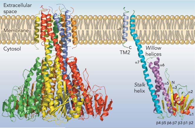
FIGURE 1. CorA Mg2+ channel.
A: CorA Mg2+ channel. The homopentamer is shown from the side with each of the five monomers in a different color. The extracellular space (periplasm) is at top. B: a single monomer is shown with TM2/α8 in gray, TM1/α7 in blue, the willow helices (α5 and α6) in purple, the β-sheet (β1-β7) in yellow, and the remaining helices (α1, α2, α3) in red.

