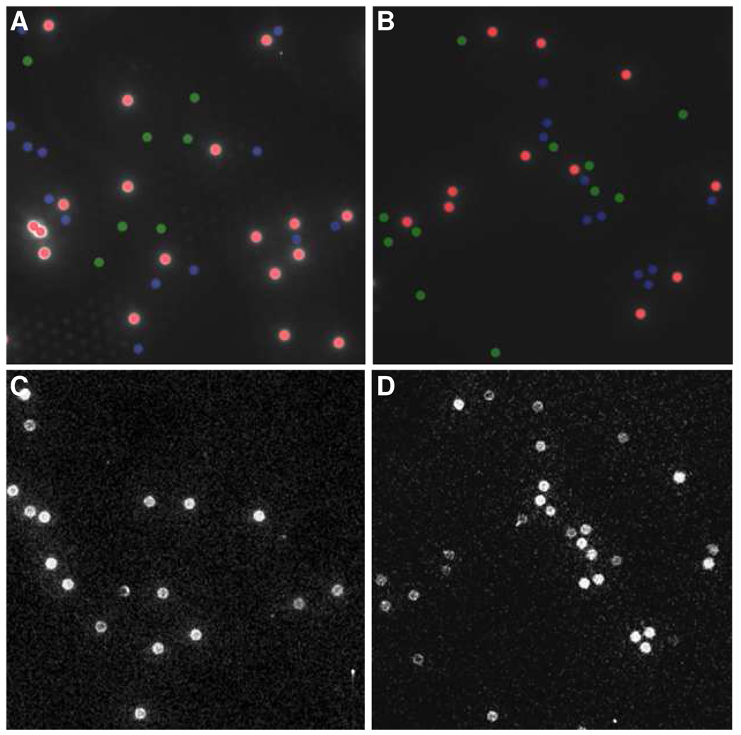
Figure 3.
(A, B) Fluorescence images of two arrays containing anti-VEGF, IL-8, and TIMP-1 modified beads (false-color overlay with color scheme same as for Figure 2). (C) ECL image of the array in (A) following exposure to a sample solution containing both [IL-8] = 50 ng/mL and [TIMP-1] = 1.5 µg/mL. (D) ECL image of the array in (B) following exposure to a sample solution containing [IL-8] = 50 ng/mL, [TIMP-1] = 1.5 µg/mL, and [VEGF] = 1.5 µg/mL

