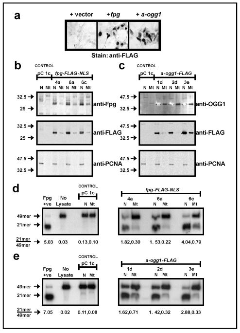
Figure 1.
Expression, subcellular localization and activity of Fpg-FLAG-NLS and α-OGG1-FLAG in stable HEK 293 clones. (a) detection of FLAG-tagged ectopic protein expression by immunocytochemistry (transient transfections). (b, c) stable expression of Fpg-FLAG-NLS (b) and α-OGG1-FLAG (c) in both nuclear (N) and mitochondrial (Mt) compartments, as observed with detection of ectopic proteins and FLAG tag by Western blotting. Membranes were re-probed for Proliferating Cell Nuclear Antigen (PCNA) to confirm purity of subcellular fractions. (d, e) in vitro 8-oxodG incision assay demonstrates increased 8-oxodG repair activity in nuclear and mitochondrial lysates from fpg (d) and α-OGG1 (e) clones. Values beneath sample lanes represent the ratio of cut (21mer) to uncut (49mer) substrate as determined by densitometry.

