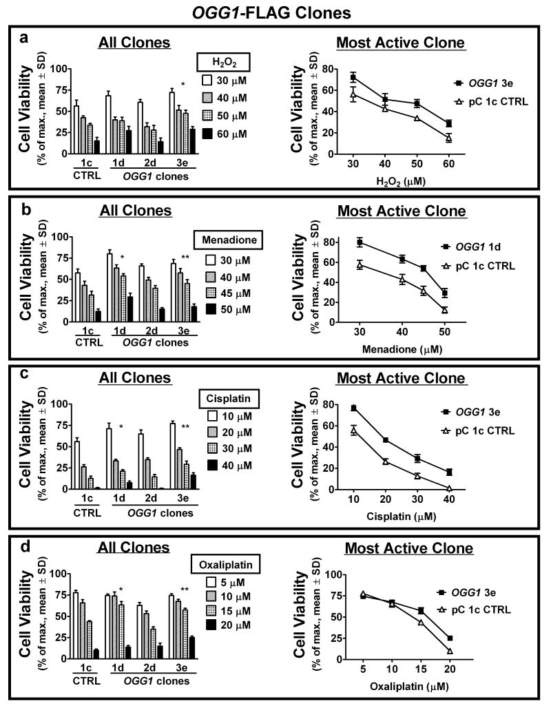
Figure 3.
Increased 8-oxodG repair via ectopic α-OGG1 expression inhibits cytotoxicity from acute exposure to ROS and ROS-inducing drugs. (a – d) cell viability of stable α-OGG1-expressing clones was compared to pC 1c control cells by colony forming assays, 7 days after one hour exposure to the indicated concentrations of H2O2 (a), menadione (b), cisplatin (c) and oxaliplatin (d). Viability response for all clones (left) and survival curve of control vs. most active clone (right) shown. Specific clones with enhanced glycosylase/AP lyase activity exhibit increased resistance to most or all treatments. *, **P<0.05 of LC50 comparable to control.

