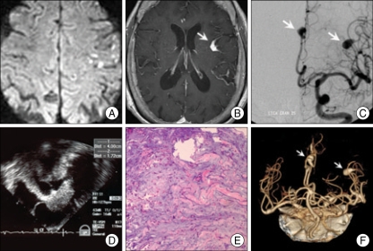
Fig. 1.
A : Multiple embolic infarctions at bilateral cerebral hemispheres on diffusion-weighted image. B : Enlarged enhanced vessel at surface of left frontal cortex (arrow) on T1-weighted image after gadolinium injection. C : Multiple fusiform aneurysms (arrows) at distal branches of anterior cerebral arteries and middle cerebral arteries on conventional cerebral angiography. D : A huge irregular shaped mass (4.0×1.7 cm) in left atrium on transesophageal echocardiograpgy. E : Prominent acid-mucopolysaccharides with interspersed round, plump and stellate-appearing mesenchymal cells on histology (H&E×100). F : No change of aneurysms (arrows) in size and number on brain computed tomographic angiography after 6 months.

