
Figure 1.
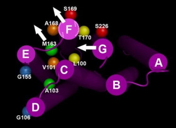
Bacteriorhodopsin model (PDB code 1C3W) viewed from the cytoplasmic side. Side chains replaced by cysteine and modified by spin labels are shown as balls at the coordinates of the respective CB atoms. Pairs of balls in the same color show double cysteine mutants to which two spin labels are attached. The three white arrows represent the possible motional reorientation of helices F and G, and the E-F loop (at Met163). The two dashed-line arrows show the possible counterclockwise rotation of helix F.