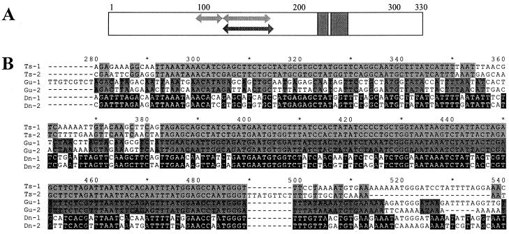
Figure 2.
Multiple alignment of Buchnera repA sequences. (A) Schematic representation of protein multiple alignment. Arrows in the region between amino acids 98 and 172 indicate location of segments that are identical among the repA1 and repA2 genes of plasmids pleu-BTs (upper arrows, positions 98–117 and 128–163) and pBGu1 (lower arrow, position 128–172). Gray blocks in the C-terminal domain indicate location and length of two deletions present in all repA2 genes. (B) Pairwise nucleotide sequence alignments of region of repA from plasmids pleu-BTs, pBGu1, and pleu-BDn, corresponding to amino acid positions 90–180, illustrating segments of complete identity in Ts-repAs (positions 292–350 and 383–489) and Gu-repAs (position 382–516).

