
Figure 5.
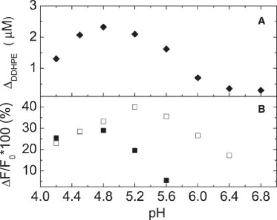
Effects of pH and His-tag on membrane binding and lipid extraction of GM2AP. (A) Plot of the change in DDHPE concentration in the supernatant as a function of solution pH for GM2AP extraction determined from sucrose-loaded vesicle sedimentation assays. For these experiments, 10 μM GM2AP was allowed to incubate for 20 min with 100 μM POPC:DDHPE (2:1) vesicles before separation by ultracentrifugation. For each pH value, the change in DDHPE concentration was referenced to samples that did not contain protein. (B) Percentage change in the fluorescence intensity detected at 518 nm with excitation wavelength of 280 nm for 10 μM GM2AP (solid) or 10 μM GM2AP10His-tag (open) with 50 μM POPC:DDHPE (9:1) vesicles as solution pH was varied. Initial fluorescence, F0, of vesicles was taken before addition of protein. The ΔF signal was determined by subtracting F0 from the value obtained (corrected for dilution) after the protein was added and allowed to incubate for 8 min. For both A and B, the solution buffers contained 25 mM NaOAc and 25 mM phosphate and the pH was adjusted by acetic acid. Each experiment was performed in triplicate and data point sizes are indicative of the error.