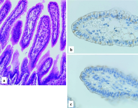
Fig. 4.
Human developing intestine, 20–21 weeks gestation. Well developed and elongated intestinal villi with occasional goblet cells (a), H-E stain ×200. Predominant cytoplasmic, but occasional membranous staining patterns of LAT1 (b), and chiefly membranous staining of 4F2hc (c), LSAB method, ×400.

