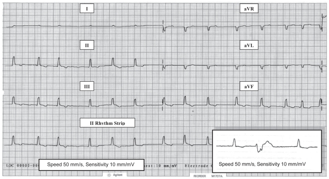
Figure 1.
Pretreatment electrocardiogram (limb leads) in right lateral recumbency from an 8-year-old male Alaskan malamute with severe hypothyroidism and dilated cardiomyopathy. There is an irregular rhythm with no visible p waves consistent with atrial fibrillation, a relatively slow rate of 125 bpm, and small ventricular complexes (R waves 0.5 mV). Inset in lead 2 (II) shows the typical PVC configuration (QS) that was seen.

