
Figure 1.
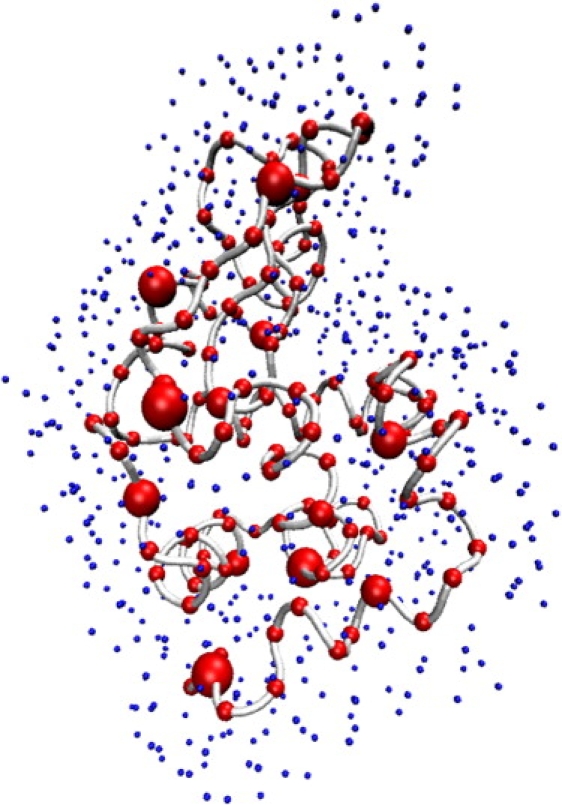
Schematic representation of lysozyme surrounded by a layer of “dummy” waters to model the local difference in relative solvent density at the surface of the protein. The scattering from a given protein conformation is conveniently and accurately represented by its N residues (red) via the Cα position, with M explicit water molecules (blue) inserted 3.5–6.5 Å away from Cα atoms to represent the hydration shell. Conformational flexibility also contributes to the scattering because of the intrinsic motions that are accessible to protein dynamics in solution.