
Figure 8.
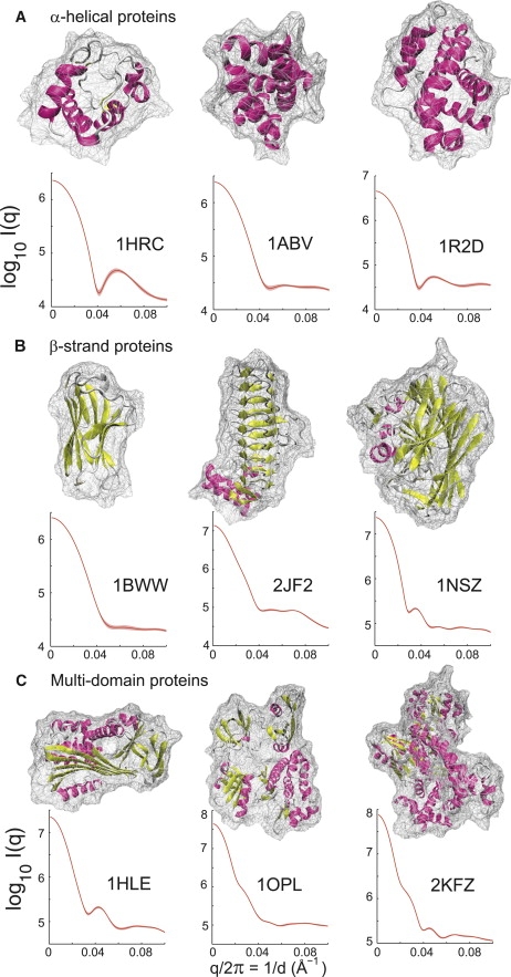
Scattering characterization of protein folds: α-helical, β-strand, and multidomain proteins. The low-angle scattering contains information about protein size and overall shape; the wider-angle scattering provides information about secondary-structure packing and domain motions. (A) The α-helical proteins: cytochrome c (PDB code: 1HRC (70)), ATPsynthase (PDB code: 1ABV (71)), and Bcl-X (PDB code: 1R2D (72)). (B) The β-strand proteins: immunoglobulin (PDB code: 1BWW (73)), acyltransferase (PDB code: 2JF2 (74)), and galactose mutarotase (PDB code: 1NSZ (75)). (C) Multidomain proteins: serpin (PDB code: 1HLE (76)), c-Abl (PDB code: 1OPL (59)), and DNA polymerase (PDB code: 2KFZ (60)). Each curve was calculated from an ensemble of snapshots taken from a 2-ns MD simulation trajectory. A log-scale of the scattering intensity was used for all the proteins.