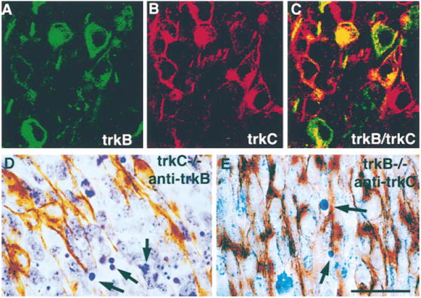
Figure 6. TrkB- and TrkC-Positive Cells Do Not Die in TrkC- and TrkB-Deficient DRGs, Respectively.

(A–C) Confocal microscopy of lumbar DRGs (middle portion of the ganglion) in a wild-type E11 embryo stained with anti-TrkB ([A], green) and anti-TrkC ([B], red) shows in a combined image that some cells express both receptors (C). Section through a lumbar DRG of a TrkC-deficient E11 embryo stained with antibodies to TrkB (D). Notice that, in spite of the presence of numerous pyknotic figures (arrows), none are positive for TrkB. Section through a lumbar DRG of a TrkB-deficient E11 embryo was stained with antibodies to TrkC (E). Only a few pyknotic profiles are observed (arrows), none of which are positive for TrkC. Scale bar, 20 μm.
