
Figure 1.
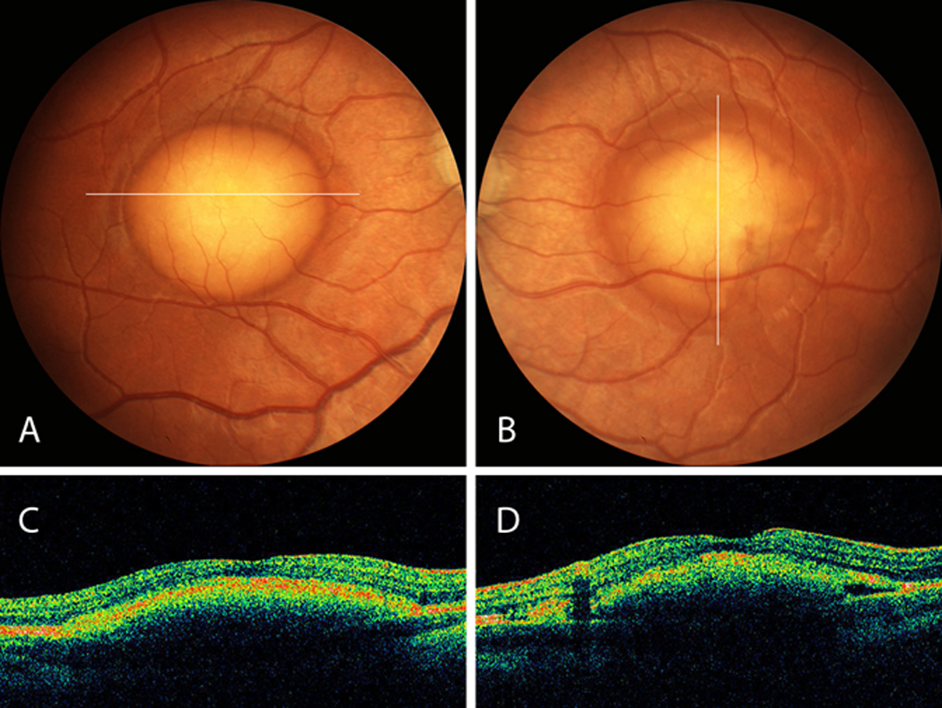
A,B, Fundus of proband 1 shows well-demarcated vitelliform lesions in the central macula. C, D, Corresponding OCT sections shows elevation at the level of the RPE with preservation of the outer retina layer.
Official websites use .gov
A
.gov website belongs to an official
government organization in the United States.
Secure .gov websites use HTTPS
A lock (
) or https:// means you've safely
connected to the .gov website. Share sensitive
information only on official, secure websites.
A,B, Fundus of proband 1 shows well-demarcated vitelliform lesions in the central macula. C, D, Corresponding OCT sections shows elevation at the level of the RPE with preservation of the outer retina layer.