
Figure 5.
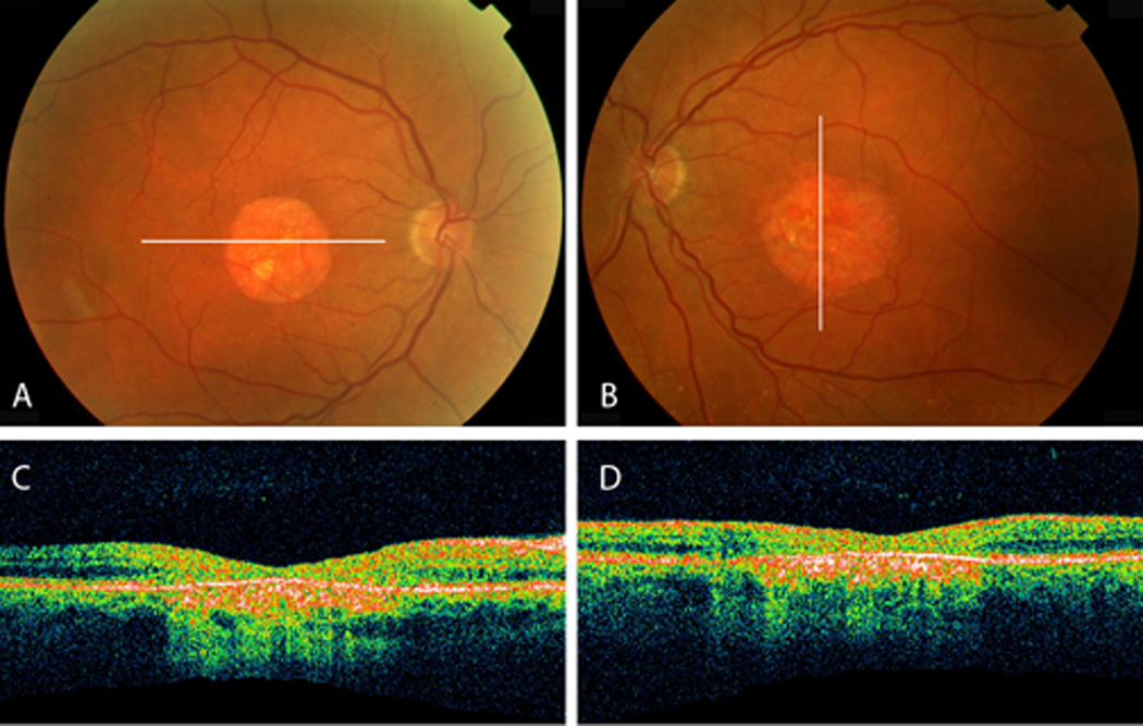
A, B, Fundus of proband 3 reveals bilateral central atrophy with few drusen in the mid-periphery of both eyes. C, D, Optical coherence tomography indicates areas of atrophy seen with increased signal posterior to the retinal pigment epithelium and attenuation of the outer retina.