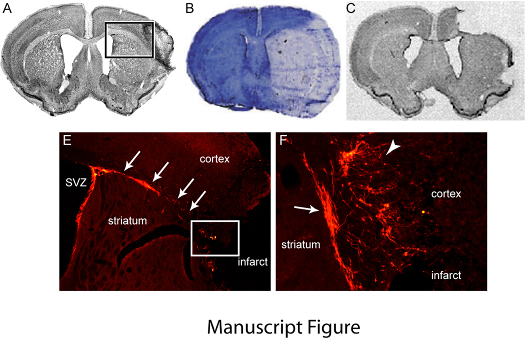
Figure. Stroke Model Determines Degree of Surviving Tissue and Pattern of Post-Stroke Neurogenesis.
A. Focal cortical stroke produced by permanent occlusion of a distal branch of the MCA and brief bilateral common carotid occlusion29. Box shows the region seen in panel D. B. Large hemispheric stroke produced by 90 minutes of MCA suture occlusion129. C. Large cortical infarct produced by permanent distal MCA and ipsilateral CCA occlusion and transient contralateral CCA occlusion130. D. Doublecortin positive immature neurons (orange) migrate from the SVZ along the white matter dorsal to the striatum to the peri-infarct cortex (arrows) at 7 days after stroke. Box shows the region that is enlarged in panel E. E. Doublecortin positive cells migrate just ventral to the infarct (arrow) and extend into peri-infarct cortex. Within peri-infarct cortex, immature neurons extend local processes (arrowhead). A comparison of the large infarcts in panels B and D indicate that the region of migration and neurogenesis in peri-infarct cortex would not be present in these stroke models as this tissue is dead. Panel B is reprinted with permission from Brain, Oxford University Press. Panel C is reprinted with permission from Journal of Cerebral Blood Flow & Metabolism, Nature Publishing Group.

