
Figure 8.
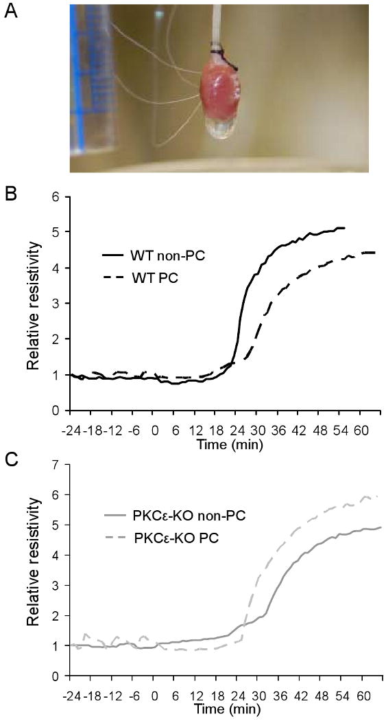
(A) Photograph showing alignment of four electrodes in perfused mouse heart for measurement of whole-tissue resistivity. (B) Representative whole-tissue resistivity curves measured in non-PC and PC hearts from WT animals. Resistivity measurements have been normalized to allow comparisons between experiments. (C) Representative whole-tissue resistivity curves measured in non-PC and PC hearts from PKCε-KO animals.