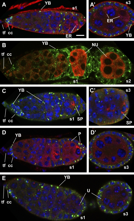
Figure 2.
Yb-containing spheres do not colocalize with known organelles in the D. melanogaster ovary. (A–E) Immunofluorescent costaining of Yb (green) and a marker (red) of the ER (A and A′), the nuage (NU; B), the sponge body (SP; C and C′), the P body (P; D and D′), or the U body (U; E) in wild-type germaria and early stage egg chambers. All of the germaria and egg chambers were oriented with the apical side to the left and the basal side to the right. tf, TF cells; cc, cap cells. s1, s2, and s3 designate stage 1, 2, and 3 egg chambers, respectively. Bar, 10 µm.

