Figure 4.
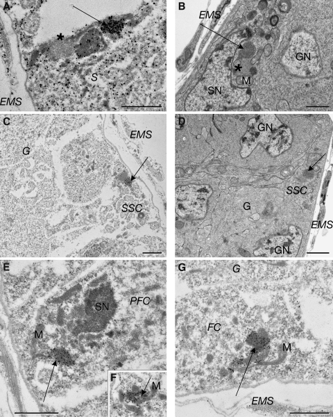
Electron microscopy of the D. melanogaster germarium revealing Yb bodies in regions II and III. (A) An immunoelectron micrograph of the frequently observed association of a Yb body (arrow) with a less electron-dense round structure (asterisk), a denser round structure, and mitochondria in somatic inner sheath cells (S) in region IIa. (B) A similar structure in an escort cell (arrow) seen by transmission electron microscopy. An adjacent gray spherical body (asterisk) presumably corresponds to the RNA-enriched spot in Fig. 1 J, based on its size, location, and association with the Yb body. (C) SSCs at the region IIa–IIb boarder show gold labeling (arrow) of a Yb body on one side of a less electron-dense structure. (D) A similar structure in an SSC (arrow) seen by transmission electron microscopy. (E and F) Immunoelectron micrographs showing that Yb bodies (arrows) in prefollicular cells are usually associated with mitochondria (M). (G) A Yb body (arrow) in follicle cells (FC) in region III associated with mitochondria. EMS, epithelial muscle sheath; G, germline cyst; GN, germline nucleus; PFC, prefollicle cell; SN, somatic nucleus. Bars, 2 µm.

