Figure 1.
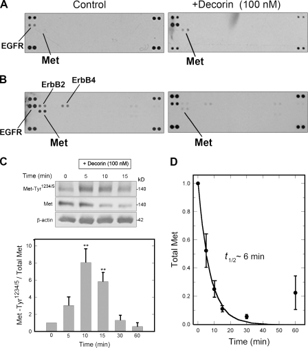
Decorin affects Met receptor signaling and turnover. (A) Phospho-RTK arrays. HeLa cells were treated with decorin for 15 min. RTK membranes were incubated with cell lysates. The duplicate dots at each corner represent phospho-Tyr positive controls. (B) The same experiment as in A using nonquiescent cells. (C, top) Representative immunoblot of a short decorin time course showing phosphorylation of the Met receptor at Tyr1234/5, total Met, and β-actin. (bottom) Quantification of immunoblots similar to those shown in the top from three independent experiments performed in triplicate. Values represent the mean ± SEM (**, P < 0.01). (D) Best-fit plot of Met receptor degradation over time. Relative values were obtained by scanning densitometry (chemiluminescence) of blots as in C and represent means ± SEM from three independent experiments performed in triplicate.

