Figure 1.
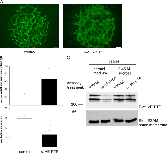
Antibodies against VE-PTP trigger vessel enlargement in allantois explants and down-regulate VE-PTP. (A) Allantois explants from E8.5 wild-type embryos were cultured on gelatin-coated ultrathin glass slides in the presence of polyclonal antibodies against VE-PTP (α-VE-PTP) or preimmune antibodies (control) for 22 h and subsequently stained by indirect immunofluorescence with a monoclonal antibody for VE-cadherin. Bar, 50 µm. (B) Average endothelial cord diameters (top) and branching points (bottom) were determined for 5 control and 5 anti–VE-PTP treated allantois explants, with 30 randomly chosen vessels per explant (as shown in A); ***, P < 0,001. (C) Confluent mouse bEnd.5 cells were treated with polyclonal antibodies against VE-PTP or preimmune antibodies for 1 h either in normal culture medium or 0.45 M sucrose containing medium (as indicated) to block endocytosis. Aliquots of cell lysates with identical protein content were immunoblotted for VE-PTP and equal loading was controlled by blotting for the endothelial antigen ESAM (as indicated on the right). Molecular weight markers are indicated on the left.

