Figure 10.
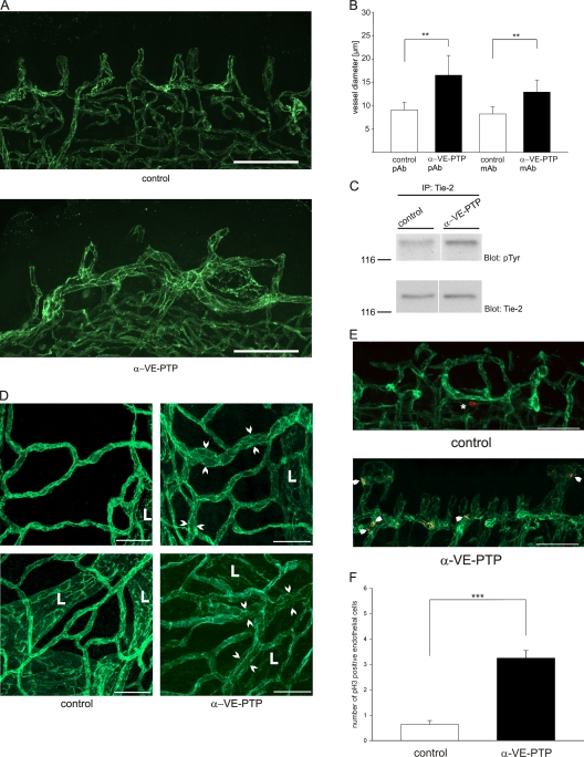
Antibodies against VE-PTP induce blood vessel enlargement and endothelial proliferation in juvenile mice and lead to activation of Tie-2. (A) 7-d-old wild-type mice were injected daily i.p. with 100 µg antibodies against VE-PTP (α-VE-PTP) or preimmune antibodies (control) for a period of 7 d. 100-µm vibratome sections of tongues were immunostained for PECAM-1. Bar, 60 µm. (B) Average vessel diameter in the tongue of wild-type mice injected daily i.p. with 100 µg polyclonal (α-VE-PTP pAb), or monoclonal (a-VE-PTP mAb) antibodies against VE-PTP, preimmune antibodies (control pAb), or monoclonal antibodies against ESAM (control mAb), respectively, for a period of 7 d. In each case three animals were analyzed. (C) 7-d-old wild-type mice were treated as described for A. Lungs of mice were lysed and subjected to immunoprecipitation for Tie-2 and subsequent immunoblotting with antibodies either against phosphorylated tyrosine or against Tie-2 (as indicated). White line indicates that intervening lanes have been spliced out. (D) Mice were treated as in A and trachea whole mounts were immunostained for PECAM-1. Arrowheads indicate enlarged blood vessels. L, lymphatic vessels. Bar, 50 µm. (E) Mice were treated for 4 d as in A and vibratome sections of tongues were immunostained for PECAM-1 and phospho-Histone 3. Asterisk, nonendothelial nucleus; arrowheads, endothelial nuclei. Bar, 50 µm. (F) Quantification of the experiment illustrated in E with phospho-Histone 3–positive endothelial nuclei counted per volume tissue (38 × 200 × 500 µm). ***, P < 0,001.

