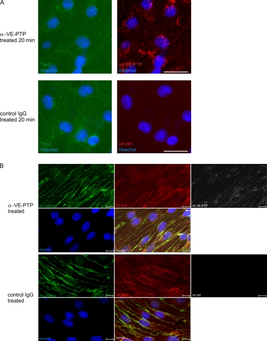
Figure 3.
Antibodies against VE-PTP do not trigger endocytosis of Tie-2 and leave VE-cadherin and VE-PTP at endothelial cell contacts unaffected. (A) Confluent bEnd.3 cells were treated with polyclonal antibodies against VE-PTP (α-VE-PTP) or preimmune antibodies (control IgG) for 20 min. Subsequently, fixed and permeabilized cells were stained with Alexa 568–conjugated secondary antibodies (internalized VE-PTP, internalized control) and for Tie-2 (Tie-2). An anti–Tie-2 staining control is shown in Fig. S5. Cell nuclei were counterstained with Hoechst. Bar, 25 µm. (B) Confluent bEnd.3 cells were treated with monoclonal antibodies against VE-PTP (α-VE-PTP) or control antibodies (control IgG) for 30 min. Subsequently, fixed and permeabilized cells were stained with Alexa 568–conjugated secondary antibodies (internalized VE-PTP, internalized control) and for VE-cadherin and VE-PTP. Cell nuclei were counterstained with Hoechst. Internalized VE-PTP was not detected with new antibodies against VE-PTP, probably because epitopes were masked by the antibodies that had triggered endocytosis. Bar, 10 µm.

