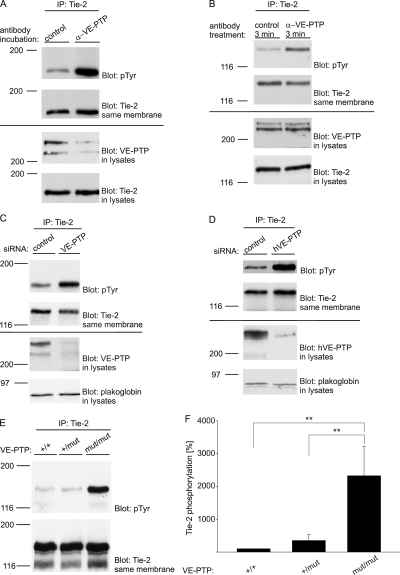
Figure 4.
VE-PTP expression inhibited by either antibodies, siRNA, or gene disruption triggers Tie-2 tyrosine phosphorylation in endothelial cells. (A) bEnd.5 cells were treated with polyclonal antibodies against VE-PTP or preimmune antibodies for 1 h and subsequently immunoprecipitated for Tie-2, followed by immunoblotting with anti-phosphotyrosine antibodies (pTyr) and antibodies against Tie-2. Aliquots of cell lysates with identical protein content were directly immunoblotted for VE-PTP and Tie-2 (bottom). (B) Similar as in A, except that antibodies were only incubated for 3 min (C) bEnd.5 cells were either transfected with control siRNA or with siRNA directed against VE-PTP. 24 h later, Tie-2 was immunoprecipitated and immunocomplexes (top) or cell lysates (bottom) were analyzed by immunoblotting with antibodies against phosphotyrosine (pTyr), Tie-2, VE-PTP, and plakoglobin (as indicated). (D) HUVECs instead of bEnd.5 cells were analyzed as in C. (E) Embryonic endothelioma cells established either from wild-type (+/+), heterozygous (+/mut), or homozygous (mut/mut) VE-PTP mutant embryos were subjected to immunoprecipitations with antibodies against Tie-2. Immunocomplexes were analyzed by immunoblotting with antibodies against phosphotyrosine or Tie-2 (as indicated). (F) Quantification of Tie-2 tyrosine phosphorylation (±SD) in wild-type (+/+; n = 2 cell lines), heterozygous (+/mut; n = 3 cell lines), or homozygous (mut/mut; n = 3 cell lines) VE-PTP mutant endothelioma, analyzed as in E. **, P < 0,01.

