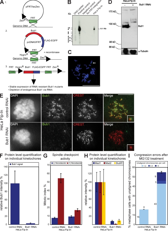
Figure 1.
Integration of the FRT site into the genome of HeLa cells does not affect the Bub1 RNAi phenotype. (A) Schematic diagram illustrating the generation of stable Flp-In and Bub1 mutant cell lines. (B) Southern blot against the DNA of two HeLa Flp-In clones probed for lacZ. The clone in the right lane was selected. The transfected vector pFRT/lacZeo was used as a positive control, and DNA from untransfected HeLa Kyoto H2B-mRED cells were used as negative control. (C) FISH analysis of selected HeLa Flp-In cells with a probe against lacZ. The arrow indicates positive FISH signal. Bar, 5 µm. (D–I) Characterization of HeLa Flp-In cells treated with control or Bub1 RNAi. (D) Immunoblot of whole cell lysates probed with Bub1 and α-tubulin antibodies. Black line indicates that intervening lanes have been spliced out. (E) Immunofluorescence images of mitotic cells stained with DAPI (DNA), Bub1 antisera (green), and CREST antisera (red, kinetochores). Bar,10 µm. (F) Quantification by immunofluorescence of Bub1 levels at kinetochores. (G) Mitotic index of HeLa Flp-In cells treated for 16 h with or without nocodazole. (H) Quantification by immunofluorescence of Mad1 (blue), Mad2 (red), and BubR1 (yellow) levels at kinetochores. (I) Cumulative plot of the percentage of metaphase cells with unaligned chromosomes after a 1-h MG132 treatment. Insets show a higher magnification view of a single kinetochore. Error bars represent standard deviation.

