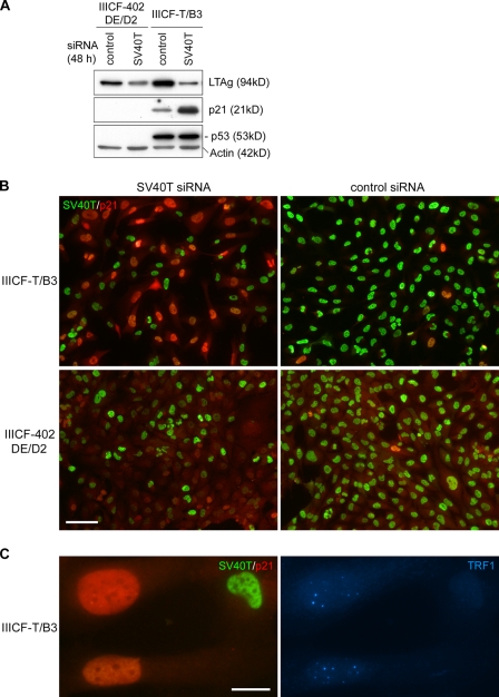
Figure 4.
Induction of APBs in SV40-immortalized ALT cells by siRNA-mediated knockdown of LTAg. (A) Western blots showed decreased LTAg in both IIICF-T/B3 (containing wt LTAg and one wt TP53 allele) and IIICF-402DE/D2 (containing mutant LTAg that does not bind p53, and no wt TP53 alleles) cells 48 h after SV40T siRNA transfection. Treatment with SV40T siRNA induced p21 in IIICF-T/B3 but not in IIICF-402DE/D2 cells. The blots were probed with the indicated antibodies. (B) Double immunofluorescence of SV40T and p21 in IIICF-T/B3 and IIICF-402DE/D2 cells treated with SV40T or control siRNAs for 4 d. Strong p21 staining was detected in IIICF-T/B3 cells depleted of SV40T. (C) IIICF-T/B3 cells were triple stained for TRF1, p21, and SV40T 4 d after SV40T siRNA transfection. APBs (visualized here as large TRF1 foci) were observed in cells with high levels of p21. Bars: (B) 100 µm; (C) 20 µm.

