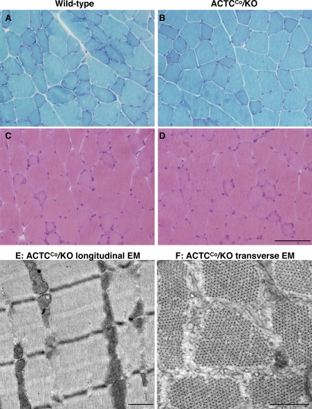
Figure 3.
Histology of skeletal muscle from male ACTCCo/KO and wild-type mice. (A–D) Representative Gomori trichrome (A and B) and hematoxylin and eosin (C and D) staining of quadriceps muscle from 3.5-mo-old ACTCCo/KO and wild-type mice. (E and F) Electron micrographs of quadriceps from 7-mo-old ACTCCo/KO mice. E shows a longitudinal and F shows a cross-sectional electron micrograph. EDL and soleus muscles were also examined and produced similar results (not depicted). Bars: (A–D) 100 µm; (E and F) 0.5 µm.

