Figure 3.
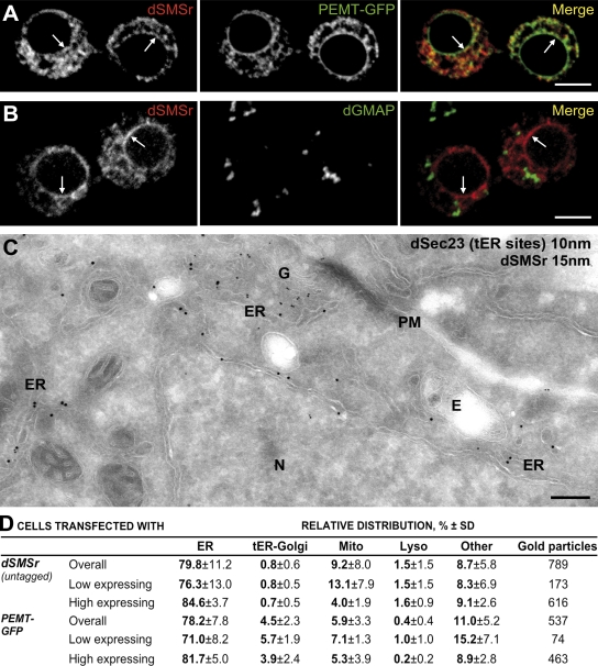
dSMSr resides in the ER of insect cells. (A) Confocal sections of Drosophila S2 cells were double transfected with native dSMSr and ER marker PEMT-GFP and immunolabeled for dSMSr. (B) Confocal sections of S2 cells were transfected with untagged dSMSr and double labeled for dSMSr and cis-Golgi marker dGMAP. (A and B) Arrows indicate nuclear envelope staining. Bars, 5 µm. (C) Localization of dSMSr by immunoelectron microscopy is shown. Ultrathin cryosections of S2 cells transfected with dSMSr were double labeled for dSMSr (15-nm gold) and dSec23 (tER site marker; 10-nm gold). G, Golgi stack; N, Nucleus; E, endosome. Bar, 500 nm. (D) Quantification of immunogold labeling of S2 cells cotransfected with dSMSr and PEMT-GFP. Low-expressing cells are those with <40 gold particles per cell section; high-expressing cells are those with ≥40 gold particles per cell section.

