Figure 3.
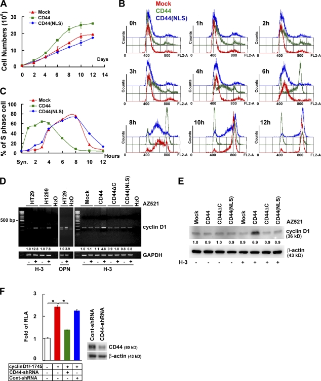
Nuclear CD44 accelerates cell proliferation and cell cycle progression through controlling cyclin D1 expression. (A) AZ521/CD44 cell clones were cultured in RPMI medium containing 1% FBS. Total viable cell numbers were determined. Data were derived from three independent experiments and are presented as means ± SD. (B and C) AZ521/CD44 cell clones were treated with H-3 and synchronized by double thymidine block. After being released into fresh medium containing 1% FBS, cellular DNA content was determined by FACS analysis after propidium iodide staining. The percentage of cells in S phase of the cell cycle is indicated in C. (D and E) Cells were cultured in serum-free medium for 24 h and incubated with or without H-3 for 1 (D) or 3 d (E). The expression of cyclin D1 was measured by RT-PCR (D) and Western blotting (E). The relative intensities of the bands, which were quantified by densitometry, are shown. GAPDH, glyceraldehyde 3-phosphate dehydrogenase. (F) Reporter assays were performed by transfection of a luciferase reporter plasmid driven by the 1.75-kb cyclin D1 promoter into H1299 cell clones stably harboring lentivirus-encoded control shRNA or shRNA targeting CD44. Data, presented as the means ± SD, were derived from at least three independent experiments. *, P < 0.05 by Student's t test. RLA, relative luciferase activity.

